We integrate smart sample preparation, and Raman analytics into a seamless workflow.
From Liters to Nanometers – Reliable Micro- & Nanoplastics Identification
Our Solution

-
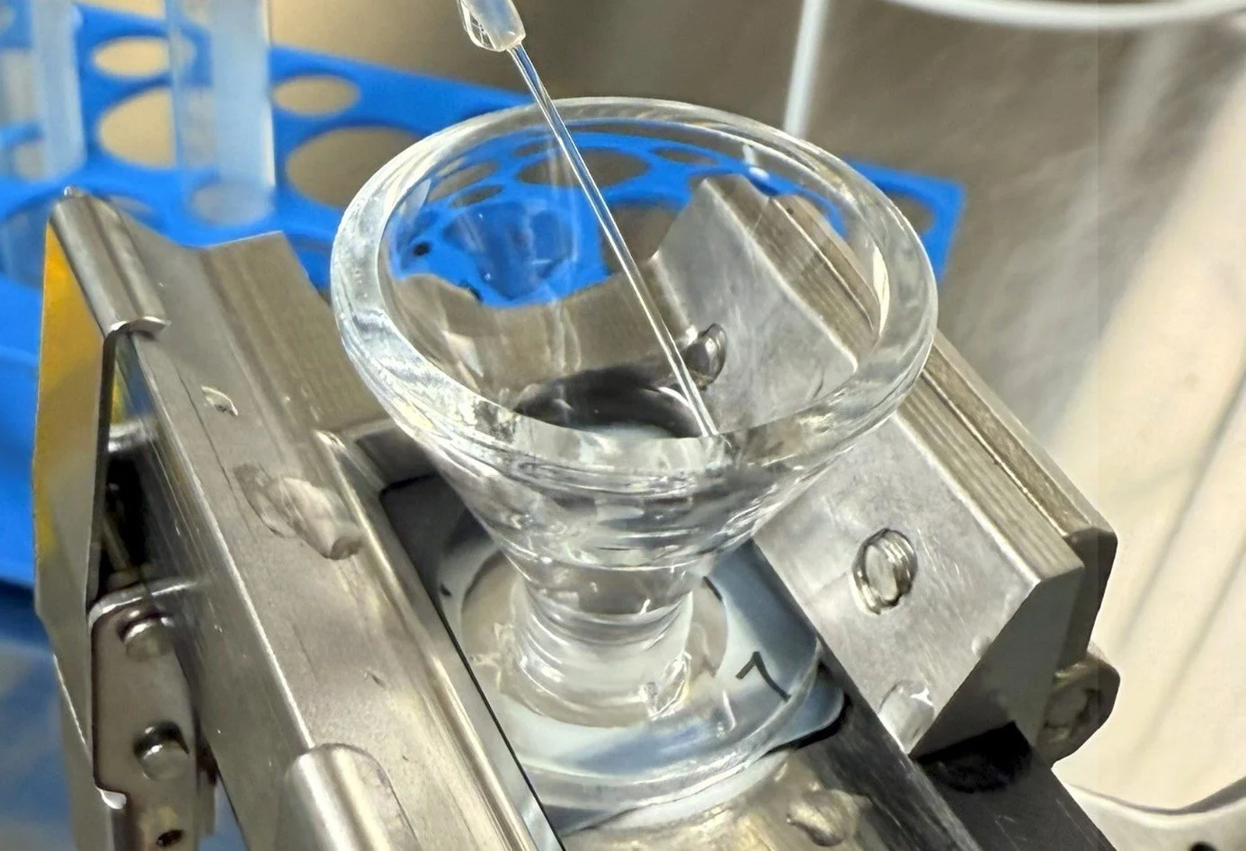
Environmental and food samples must be concentrated from liters of liquid down to milliliters.
-
Samples contain not only plastics, but also fibers, minerals, and organic substances.
-
From 500 nm up to 5 mm – covered by software controlled magnification in light microscopy. This allows reliable detection of both submicron fragments and larger particles.

Our Workflow
-
Density separation, enzymatic and oxidative pretreatment
Low-background specialty filters for clean deposition
-
Homogeneous particle distribution across the filter surface
Reduced overlap → higher detection confidence
-
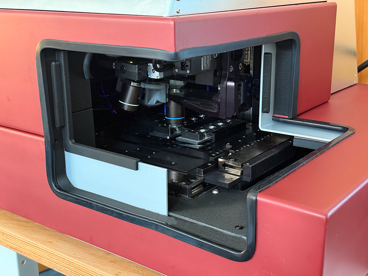
DeepMorph detects and classifies particles by size & morphology
RamanMetrix identifies polymers via spectral fingerprints
Autofocus and scanning illumination guarantee sharp imaging across the entire filter surface
Advanced thresholding algorithms ensure homogeneous and comparable particle counts and morphology results
Sensitivity down to 500 nm
-
Statistics: counts, morphology, polymer types
Robust through PCA and Pearson correlation
Reports authored and supervised by experienced scientists
Based on a vast library of neutral and colored plastics across different stages
Machine learning can be tuned to trace specific sources in manufacturing and filling processes (e.g. packaging, raw material handling, closures)
Auditable reports under ISO 9001 QMS
Raman Spectroscopy: Reading the Fingerprint of Plastics
Every polymer has a unique vibration pattern of its chemical bonds.
Raman spectroscopy reveals these vibrations – like a spectral fingerprint.
Each plastic type (e.g., PET, PE, PS) has a distinct Raman signature.
Even colorless or highly fragmented particles can be identified with confidence.
Combining morphology with chemistry provides a complete picture:
→ Yes, it’s plastic – and here’s the type.
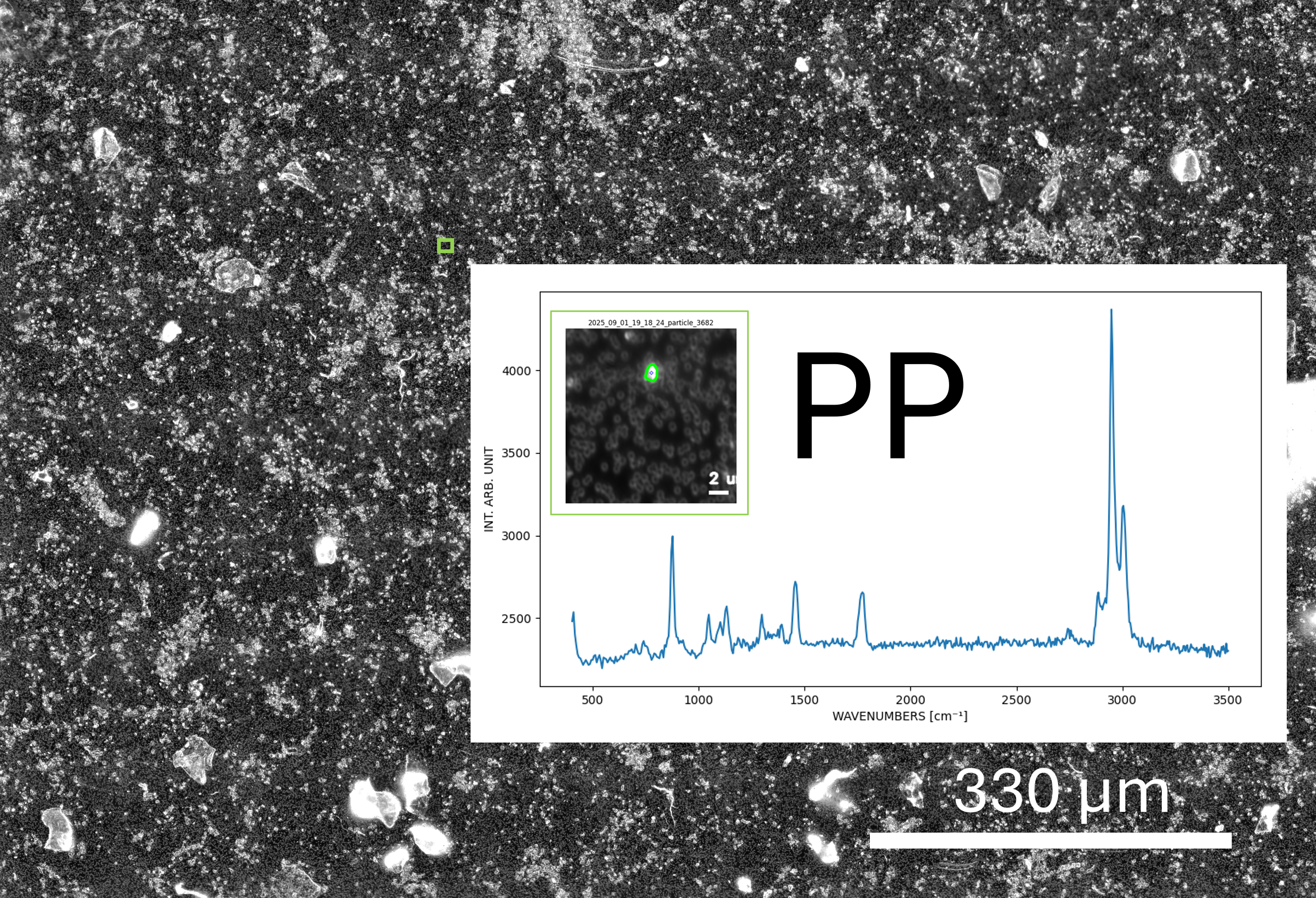
Raw data, from scanning to reliable Raman signatures of submicron particles

Why Raman instead of FTIR?
Higher spatial resolution: Raman achieves submicron focus, while FTIR is typically limited to >10 µm.
Dry samples, negligible water interference: Unlike FTIR, Raman analysis is not compromised by strong absorption bands from water — an advantage for both dry and aqueous-origin samples.
Speed & automation: Raman integrates seamlessly with automated imaging, autofocus, and AI-driven classification.
Fluorescence challenge — but manageable: Some particles emit fluorescence that can mask the Raman signal. In practice, fluorescence is usually caused by contaminants rather than the polymer itself. The smaller the fragment, the less fluorescence tends to occur — which makes Raman particularly strong in the submicron regime.
Future-ready: With advanced optics and algorithms, Raman is the most promising path to extend detection below 500 nm, enabling robust nanoplastics analysis.